It’s been awhile since I posted any needlework images. Here is a smattering of pieces made in the last year or two…
FLOUNCE SLEEVE
 |
| Flounce Sleeve. Artist: Kriota Willberg |
The late great Rozsika Parker, in her fabulous book, The Subversive Stitch, described the way Enlightenment philosophers and scientists introduced the notion that women were essentially biologically driven to sew. At the same time, women were physically associated with flowers in some anatomical texts by Casseri and Van de Speigel. I decided to up the fantasy a notch by re-designing a flounce sleeve embroidery pattern from an 1855 issue of Godey’s Lady’s Book using anatomical images from The surgery, surgical pathology and surgical anatomy of the female pelvic organs : in a series of coloured plates taken from nature, with commentaries, notes and cases,by Henry Savage (1876). You can find this book at the New York Academy of Medicine Historical Collection.
I worked up the pattern in cotton floss on linen. Then I accidentally burnt it with an iron when I had a migraine. It took two months to make.
 |
| Oophorectomy. Try saying that 5 times, fast! Artist: Kriota Willberg |
OOPHORECTOMY
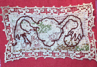
An oophorectomy is a surgery where an ovary is removed. This is a free-hand image of a (somewhat cartoonish) uterus, fallopian tubes, and one ovary. I darned yarn into a torn lace…doily? Then stitched the uterus in darker yarn. I imagine the darned doily as a representation of the broad ligament. I pulled and puckered the yarn in the area of the missing ovary to represent scarring that might occur after an oophorectomy.
CATGUT TISSUE SLIDE
 |
| Tissue slide cross stitch pattern made in Photoshop. Kriota Willberg |
 |
| Catgut tissue slide cross stitch. There's a typo in there! I'm not saying where- find it yourself! Artist: Kriota Willberg |
I thought you might be curious as to what a pattern for one of my cross stitches looks like.
The top image is a counted cross-stitch pattern, assembled in photoshop. It's a photomicrograph of catgut imbedded in dog tissue from Textbook on Sutures(1942) by Paul F. Ziegler, and is supplemented by images from The Gentleman’s Dog, his Rearing, Training, and Treatment(1909) by C.A. Bryce, and Trichologia mammalium; or, A treatise on the organization, properties and uses of hair and wool, together with an essay upon the raising and breeding of sheep. You can access these books at the Academy Historical Collection. The text forming the border of the images is liberally excerpted from the textbook on sutures and says,
“…catgut is made from the first 6-8 yards of the stomach-end of the small intestine of sheep. The absorption rates of catgut sutures are regularly checked by suture implantations in muscle of laboratory animals (dogs and rabbits). Following implantation, the surrounding tissue reacts to wall off and digest this foreign body. A disintegration occurs, small fragments are phagocytosed by macrophages and are thus digested.” The delineation of the dog and sheep image have been redrawn to mimic clumps of macrophages, which white blood cells.
The catgut and macrophages are made with cross-stitch. The dog tissue is tinted by using a half tent stitch, and the areas absent of stitching are the forming granulation tissue.
This piece took about 4 months to make! (Okay, yeah, because I had to work and do other things at the same time, but still!)
MEDICAL IMAGERY THROUGH EMBROIDERY WORKSHOP
This spring (2019) I traveled up to the Rochester Institute of Technology, spoke about graphic medicine, and lead workshops on injury prevention and Medical Imagery through Embroidery! Yup! The workshop was a blast. I presented a slide show on cultural and aesthetic messages that we can interpret from educational anatomical and medical imagery. We discussed anatomical symbolism used by artists in their non-medical work. Then I introduced the students (from the medical illustration, art, and English departments) to some basic embroidery stitches, gave them fabric with pre-printed historical anatomical images, embroidery supplies, fabric pens, and fabric, and let them transform the “academic” “professional” imagery into something more personal. They did some great work! I don’t have any examples of student work, but I can show you a piece I worked up using the image of a child’s skull from William Cheselden’s Osteographia, fabric pens, tulle, and embroidery.
TOM’S TUMOR AND THYMUS
My friend Tom had a thymectomy. His surgeon took a photo of Tom’s thymus with it’s (benign!) tumors and gave it to him. Tom knew I’d love to turn it into a needlepoint piece, so he gave me a copy of the photo and permission to work it up. I made it on monocanvas with wool yarn. One thing I like about many decorative projects is the way they label or name their subjects in the context of the piece, be it a painting, needlepoint, or a tattoo. So I did it too.
 |
| Tom's Tumor and Thymus. Artist: Kriota Willberg |
This is not Tom's favorite of my work. (He is a little squeamish about it. Who can blame him?)

No comments:
Post a Comment